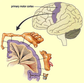
Primary motor cortex
From Psy3242
== Structure and Function == 
The primary motor cortex, also known as the motor strip, M1, F1, or Brodmann's Area 4, is one of the principal brain areas responsible for motor function and the execution of movements in muscles. It is located in the rear portion of the frontal lobe of the brain, before the central sulcus that separates the frontal lobe from the parietal lobe and along a bump called the precentral gyrus. M1 is only part of the complete motor cortex, which is also made up of the premotor area (PMA) and the supplementary motor area (SMA) of Area 6, but it includes the cortico-spinal and the cortico-bulbar pathways and the axons of pyramidal neurons.
The motor strip is highly topographical in its organization, and every part of the body is represented, though the relationship between cortical 'space' in the motor strip and body region is not proportionate. Body areas capable of fine motor control - such as the hands and fingers and the mouth area of the face - are over-represented, and there is under-representation of less 'movement-critical' regions such as the back and top of the head, the trunk or torso of the body, and the upper limbs. The amount of brain matter in M1 devoted to each particular body part represents the amount of control that it has over that body part. The disproportionate nature of the body's representation is displayed in a map called the motor homunculus (featured below).
The primary motor cortex is predominantly contralateral in its control over the body. The right motor cortex coordinates and manages muscles in the left side of the body, and the left motor cortex coordinates the right side of the body. Signals from M1 cross the body's midline to activate skeletal muscles on the opposite side of the body.

The Cortico-Spinal Tract
Neurons in M1, the SMA, and the PMA constitute the fibers of the cortico-spinal tract, the main pathway for the control of voluntary movements in humans. Composed of millions of these fibers, it is the only direct pathway from the cortex to the spine and descends through the brain stem where most of the fibers branch over to the opposite sides of the body. The fibers then continue down the spinal cord until they reach their appropriate levels or locations.

History
In 1870, Hitzig and Fritsch electrically stimulated different parts of a dog's motor cortex. They observed that depending on where they applied stimulation, various parts of the body contracted. When they destroyed the same small area of the cortex, the dog experienced permanent paralyzation in the corresponding body part. Hitzig and Fritsch became the first to come to the conclusion that every part of the body has a particular region of the primary motor cortex devoted to its movement.
The location of the primary motor cortex was further confirmed in the mid-20th century during brain operations conducted by neurosurgeons such as Dr. Wilder Penfield. Penfield stimulated different areas of the cortex during a surgery on an epileptic patient to identify the vital regions not to be removed surgically. In doing so, Penfield realized there was a somatotopic representation of corresponding body parts in the primary motor cortex.
Interesting fact:
The motor cortex is responsible for movement, foot tapping, dancing and playing an instrument while the auditory cortex is responsible for the first stages of listening to sounds, and the perception and analysis of tones.
Damage and Disorders
Extensive damage to the area of the primary motor cortex can result in a widespread loss of muscle function and paralysis. In some cases in which the entire left or right primary motor cortex has been damaged, lasting impairments including hemiplegia can occur. Causes can include an aneurysm, hemorrhage, blood clot, an accidental head injury, epilepsy, or a tumor. An individual affected by hemiplegia is no longer able to intentionally move parts of their body on the side opposite to that of the brain damage. However, there is usually a discernible degree of recovery of function over time as the blood supply to neurons adjacent to the damaged areas normalizes.
Another disorder resulting from trauma to the primary motor cortex during fetal development or birth is called cerebral palsy. The disorder varies across individuals but a hallmark is motor disturbance, which may include poor muscle coordination, unwanted involuntary movements, and excessively tensed muscles.
