Wernicke's area
From Psy3241
Kgutekunst (Talk | contribs) |
Kgutekunst (Talk | contribs) (→References) |
||
| Line 10: | Line 10: | ||
| - | Image taken from: | + | Image taken from: |
| + | http://users.fmrib.ox.ac.uk/~stuart/thesis/chapter_3/image3_15.gif | ||
Revision as of 17:07, 23 April 2008

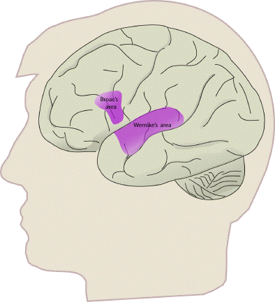
Wernicke's area was discovered by Karl Wernicke in 1874. It is the left superior temporal gyrus. Specifically, this area controls the function of connects speech sounds to stored representations of words. Wernicke's area is anatomically linked to Broca's area. A lesion to this area will likely result in difficulties in language comprehension. In an article published by Carl Wernicke in 1874, he reported 10 aphasic patients with difficulties in language comprehension. An autopsy on four of the patients provided results that they had lesions damaging the left temporal lobe. This specfic type of aphasia is now known as Wernicke's aphasia. Recent research by Dronkers et. al. (1998) revealed that 'pure' damage to Wernicke's area most likely results in impairment in repetition rather than comprehension deficits. Damage to the part of the brain linking Broca's area and Wernicke's area, the arcuate fasciculus, can lead to conduction aphasia, in which the patient loses the ability to repeat words.
Other brain areas associated with language function include: Broca's area, supramarginal gyrus, angular gyrus, and arcuate fasciculus.
References
Image taken from: