Eye
From Iusmhistology
Revision as of 17:47, 12 April 2011 by 134.68.138.106 (Talk)
- started here on 04/11/11.
Contents |
The eye
Optical anatomy
- Important anatomical features of the eyeball include:
- cornea with limbus (outer rim that holds stem cells)
- anterior chamber
- pupil
- iris
- posterior chamber
- ciliary body
- lens (with ciliary processes and zonules)
- vitreous body
- retina
- ora serrata
- fovea
- optical disc

Wall of the eye
- The wall of the eye is comprised of three layers: fibrous tunic, uvea, retina (where the retina is the inner-most layer).
- The fibrous tunic generates the cornea, the sclera, and provides continuity with the conjuctiva.
- The uvea is the vascular tunic (tunica vasculosa) that carries the vasculature of the eye.
- The uvea also makes the iris, ciliary bodies, and the choroid.
- The choroid is highly vascularized.
- The retina provides the sensory material of the eye.
- The retina includes the pigmented epithelium, the neural photoreceptor cells, and the neuronal integreative circuitry and supporting cells.
- Recall that photoreceptor cells come in rods and cones.
- Note that the pigmented epithelium is deep to the photoreceptors.

Eyeball function by component

Cornea
- The cornea is the most superficial layer of the eye and is exposed to the environment.
- Recall that the cornea arises from the fibrous tunic of the optical tissue.
- The cornea is transparent, avascular, and bends the incoming light to hit the lens.
- In fact the cornea bends the light (refracts) even more than the lens.
- Photorefractive keratectomy and LASIK (laser in situ keratomileusis) are two surgeries that reshape the deeper (non-epithelial) layers of the cornea to correct poor refraction that results in poor vision.
- The cornea has three cellular layers and two atypical basement membranes.
- The layers (superficial to deep): epidermis, Bowman's membrane, stroma, Descemet's membrane, and endothelium.
- With age, Descemet's membrane decreases in transparency and leads to decreased light transmission.

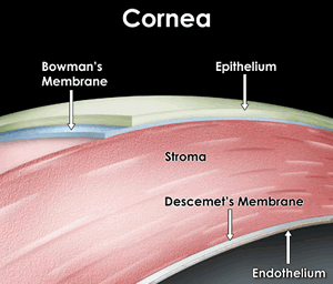


Epithelium of the cornea
- The epithelium of the cornea is made of stratified, squamous, non-keratinized epithelium.
- The epithelium layer is highly proliferative and has abundant pain fibers.
- The epithelial layer maintains a population of stem cells in the limbus area (the junction of the cornea with the sclera).
- Note that aqueous humor (from the anterior chamber) drains through the corneoscleral junction (the limbus).
- We call the corneoslceral junction (the limbus) a trabecular meshwork and the site of Schlemm's canal.


Stroma of the cornea
- The stroma is also called the substantia propria.
- There are 50-60 layers of cells making the stroma the most substr
- The stroma is series of layers of fibrocytes, proteoglycans, and ECM fibers with alternately oriented collagen fibers.
- Note that collagen of the stroma is of type 1 and 5 and are non-fibrillar.
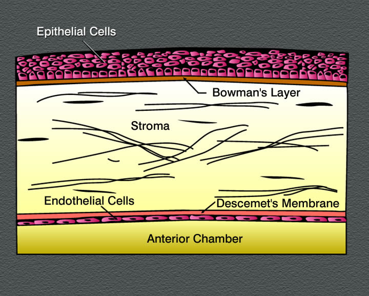
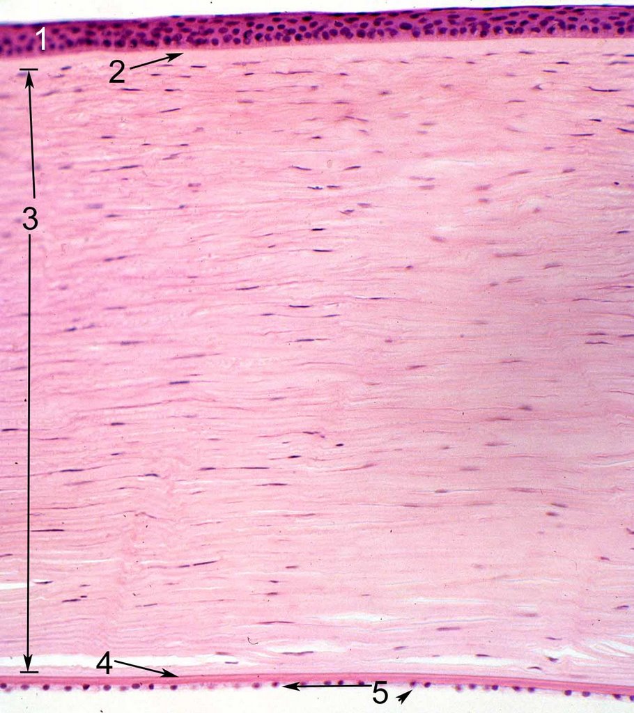

Endothelium of the cornea
- The endothelium of the cornea contains leaky intercellular junctions that allow exchange of fluid with the anterior chamber.
- Drainage by the endothelium keeps the stroma relatively dry.
- Note that fluid transport by the endothelium contributes to the transparency of the cornea.


Uvea
- Recall that the uvea is one of the three layers of the eye: the tunica fibrous, the uvea, and the retina.
- Recall that the uvea is also called the tunica vasculosa and carriers the vasculature of the eye.
- The uvea includes the iris, the ciliary bodies, and the choroid.
- Just as the cornea is the anterior portion of the fibrous tunic and is completed posteriorly by the sclera, the iris and ciliary body are the anterior aspect of the tunica vasculosa and are completed posteriorly by the choroid.

Choroid of the uvea
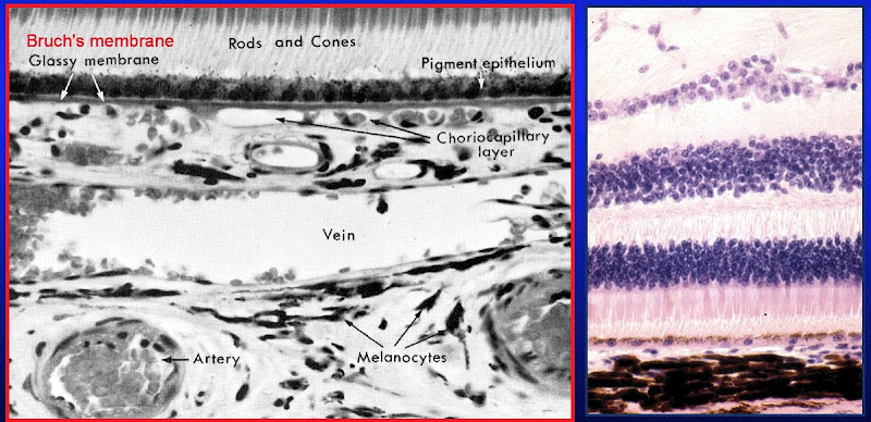
- Recall that the choroid is the posterio-lateral aspect of the uvea (tunica vasculosa).
- The choroid carries vasculature to the retina and the sclera.
- The choroid is highly pigmented and found between the sclera (part of the tunica fibrous) and the retina.
- The choroid has three layers: sclera vasculature, retinal vasculature, and Brunch's membrane.
- Note that these layers are indistinguishable in the lab.
- The choriod layer is separated from the retina by the Bruch's membrane; that is, Brunch's membrane is the deepest aspect of the choroid part of the tunic vasculosa (uvea).

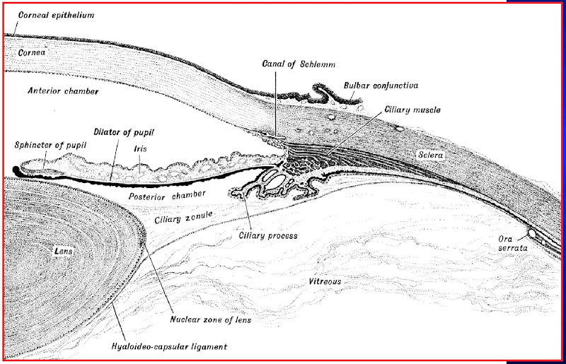
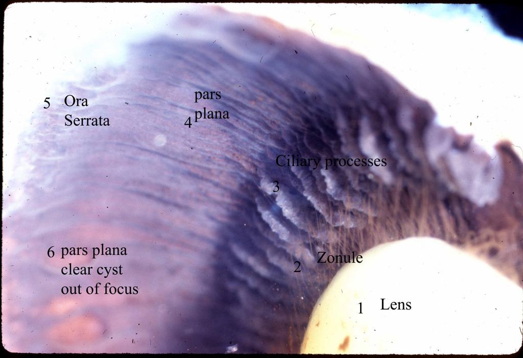
Ciliary body
- The ciliary body is a thickening of the vascular layer near the anterior aspect, radially from the lens.
- Ciliary bodies have two structures: ciliary zonules (ligaments) and ciliary processes.
- Ciliary zonules (suspensory ligaments):
- Ciliary ligaments project from the tunica vasculosa to the lens capsule and control the flattness / bulging of the lens in order to focus light (a process called accomodation.
- Upon contraction of the ciliary muscle, the many ciliary zonules are brought closer together and anterior which actually allows the lens to relax.
- Far vision requires a flattened lens (think flat like a frisbee which you hope will go "far") so the ciliary muscle relaxes, the zonules tense, and the lens is pulled into a flatter shape.
- Near vision requires a bulged lens so the ciliary muscle contracts, the zonules relax, and the lens relaxes into a bulge.
- Recall too that near vision gives eye strain which is the excessive contraction of the ciliary muscles.
- Oxytalin fibers are used to attach the basement membrane of the non-pigmented epithelium of the ciliary body (part of the uvea) to the basement membrane (called the lens capsule) of the lens.

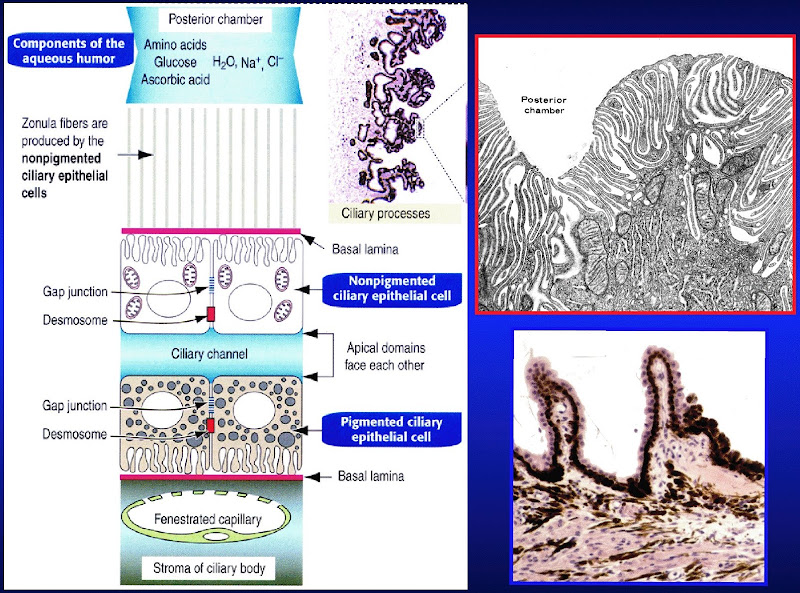
- Ciliary processes:
- Ciliary processes primarily function to generate aqueous solution.
- Aqueous solution provides nutrition to the lens and maintains intraocular pressure.
- Aqueous solution has little protein, some glucose, and similar ion concentration as plasma.
- Ciliary processes are an extension of the tunica vasculosa (the uvea) and are highly vascularized.
- There are two layers to the ciliary process tissue: deep layer (pigmented) and surface layer (secretory, non-pigmented).
- The pigmented layer (deep layer) of the ciliary process is continuous with the pigmented layer of the retina.
- The non-pigmented layer's (secretory, surface layer) apical surface faces the pigmented layer and the basolateral surface has lots of folds and borders the posterior chamber.
- Blood-aqueous barrier: occluding junctions at the apex of the ciliary process's surface epithelium keeps blood and aqueous solution from mixing.
- Note that glaucoma results from blocked drainage of aqueous fluid and the resulting elevation of intraoccular pressure.
- Ciliary processes primarily function to generate aqueous solution.

.jpg)
Iris
- The iris the most anterior aspect of the uvea (the vascular layer).
- Recall that the uvea is the middle layer of the three layers of the eyeball: tunica fibrous, tunica vasulosa (uvea), and the retina.
- The iris divides the eyeball into two chambers: the anterior and posterior chambers.
- The iris's job is to regulate the amount of light entering the eye ball.
- The iris is primarily composed of stroma with an epithelial covering only on the posterior surface.
- The stroma is highly vascular (recall that the iris is part of the tunica vasculosa) and has fibroblasts, connective tissue, melanocytes, and myocytes.
- It makes sense that the stroma of the iris has melanocytes and myocytes because the iris provides our eye color and opens and closes to regulate light.
- The posterior aspect of the iris has epithelial and myoepithelial layers that provide the pigment of eye color and the movement of the iris, respectively.
- Note that the stroma contains the constrictor muscle of the pupil and the myoepithelium contains the dilator muscle of the pupil.
Eye color
- Eye color is determined by the quantity and distribution of pigment cells in the stroma and posterior epithelilum of the iris.
- Pink: complete lack of pigment; albinism (lack of tyrosinase or a defect in melanosome transport, RAf27a)
- Blue < grey / green < brown => less to more pigment.
